Introduction
X-ray Technology, a pivotal clinical development, has been an essential device in the field of medical services for an extended period. Since its disclosure 1895 by Wilhelm Conrad Roentgen, X-beams have upset clinical diagnostics, offering harmless bits of knowledge into the human body.
The Verifiable Excursion of X-ray Technology
The historical backdrop of X-ray Technology is a wonderful one, set apart by luck, development, and quick headway. Everything started in 1895 when Wilhelm Roentgen, a German physicist, made an unplanned revelation. While exploring different avenues regarding cathode beams, he saw that a screen in his research facility began to shine even though it needed to be more straightforwardly presented to the cathode beams. Intriguingly, the wellspring of this strange sparkle was a piece of barium platinocyanide put a few feet from the cathode beam tube. Roentgen named this baffling peculiarity “X-beams,” with the “X” connoting their obscure nature.
Roentgen’s revelation created an uproar among established researchers and then some. The capability of X-beams as a demonstrative device became apparent, and in a year, X-beam machines were being utilized in clinical settings. The principal X-beam picture, a radiograph of Roentgen’s better half’s hand, established the groundwork for a transformation in clinical imaging. It wasn’t some time before X-beams were utilized to envision cracks, unfamiliar articles, and structures inside the human body.
Early X-beam machines were simple, contrasted with the present complex gadgets. They presented patients with higher radiation dosages and needed more accuracy and clearness. We currently partner with X-beam imaging. Even so, they prepared for the advancement of this innovation.
The Coming of Current X-ray Technology
The advancement of X-ray Technology in the mid-twentieth century saw massive enhancements in hardware, security measures, and picture quality. Developments, for example, the Coolidge tube, which was considered more controlled and proficient X-beam creation, and the presentation of lead covers and collimators for radiation insurance, changed X-beam imaging into a more secure and more exact symptomatic instrument.
During the mid-twentieth 100 years, radiography saw significant progressions, including the acquaintance of difference specialists with upgraded delicate tissue representation. These specialists, like barium and iodine, assumed an urgent part in concluding different ailments.
Computerized Radiography
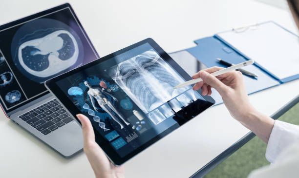
Yet again, the computerized upset of the late twentieth century changed X-ray Technology. Customary film-based radiography gave way to advanced radiography (DR) and figured radiography (CR), offering a few benefits. Computerized X-beam pictures could be procured, handled, and put away electronically, making them effectively open and considering post-handling to upgrade picture quality. This progress additionally diminished the utilization of unsafe synthetic compounds and the requirement for actual film stockpiling.
The approach of PACS (Picture Chronicling and Correspondence Framework) permitted medical services experts to access and share X-beam pictures from a distance, working with convenient finding and discussion. Also, the mix of artificial brainpower (computer-based intelligence) in radiology has upgraded the translation and examination of X-beam pictures, empowering speedier and more precise judgments.
Utilizations of X-ray Technology in Medical Services
X-ray Technology has a large number of utilizations in medical services, stretching out past conventional radiography. A portion of the critical regions where X-beams are used include:
Radiography: Conventional X-beam radiography stays a foundation of clinical imaging. It is utilized to distinguish cracks, disengagements, and bone-related pathologies.
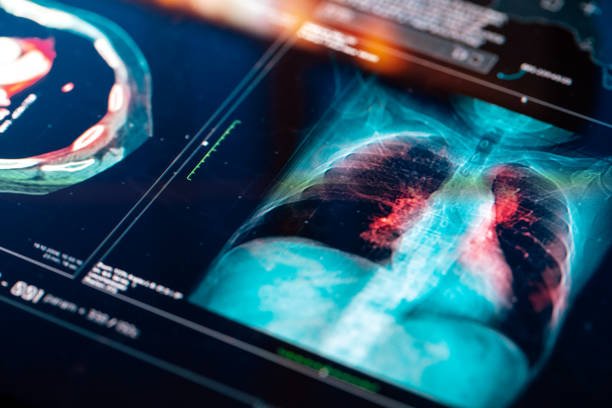
Fluoroscopy: This ongoing X-beam imaging strategy is utilized in barium swallow studies, angiography, and cardiovascular catheterization to picture dynamic cycles inside the body.
Mammography: X-beam mammography is pivotal for bosom malignant growth screening and early discovery, possibly saving innumerable lives.
Interventional Radiology: X-beam-directed methodologies, like angioplasty, embolization, and stent arrangement, are used in interventional radiology to treat different circumstances without requiring intrusive medical procedures.
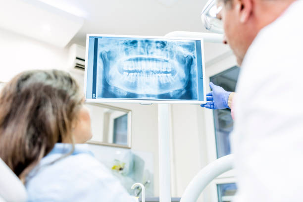
Dental Radiography: X-beams are broadly utilized in dentistry for diagnosing dental caries, oral contaminations, and jaw issues.
Veterinary Medication: X-ray Technology isn’t restricted to human medical services. It is widely utilized in veterinary medication for diagnosing and treating different creature conditions.
The Effect of X-ray Technology on Medical Care
The effect of X-ray Technology on medical services is limitless. It has achieved a considerable number of advantages, both for patients and medical services suppliers:
Early Determination: X-ray Technology empowers the early recognition of sicknesses and conditions, fundamentally working on persistent results. For instance, recognizing cracks or cancers in their underlying stages is considered a more viable therapy choice.
Non-Intrusive Imaging: X-ray Technology is harmless, decreasing patient inconvenience and confusion related to obtrusive systems.
Precision and Exactness: With computerized radiography and high-level imaging methods, X-ray Technology offers high-goal pictures that improve demonstrative precision.
Efficiency: X-ray Technology speeds up the symptomatic cycle, empowering medical services suppliers to go with reasonable therapy choices.
Reduced Radiation Openness: Progressing endeavors to diminish radiation dosages in X-beam systems have prompted more secure imaging works, limiting the potential well-being gambles related to radiation openness.
Research and Improvement: X-ray Technology keeps on driving clinical examination and development. It helps with the improvement of new analytic apparatuses and treatment modalities.
Education and Preparing: X-ray Technology is essential in clinical schooling and preparing, assisting future medical services experts with acquiring fundamental abilities in picture understanding.
Difficulties and Future Turns of Events
While X-ray Technology has made some fantastic progress, it faces difficulties. Radiation openness remains a worry, especially for continuous and high-portion applications. Finding some harmony between the advantages of X-beam imaging and radiation security is a constant undertaking.
Also, the coordination of artificial intelligence in radiology is ready to upset X-beam understanding further. Simulated intelligence calculations can help radiologists recognize irregularities and make more exact conclusions, eventually working on persistent consideration.
The improvement of more current imaging methods and hardware, for example, 3D X-beam imaging and versatile X-beam gadgets, keeps increasing the abilities of X-ray Technology. These developments vow to improve the accuracy and extent of X-beam diagnostics before long.
Conclusions
X-ray Technology has traveled from an inadvertent disclosure to a foundation of current medical care. Its effect on quiet consideration, sickness analysis, and clinical exploration is incomprehensible. The advancement of X-ray Technology from its initial, unrefined structure to the present modern computerized radiography and simulated intelligence helped to understand and change the area of radiology.
As innovation keeps propelling, X-beam imaging will stay at the front of indicative medication, giving essential bits of knowledge into the human body. The continuous quest for more secure and more effective imaging strategies will guarantee that X-ray Technology will keep saving lives and add to the headway of medical care for a long time.

FAQS
What is X-ray Technology?
X-ray Technology includes the utilization of X-beams, a type of electromagnetic radiation, to take pictures of the human body and different items. These are called X-beam pictures or radiographs and are fundamental for clinical findings and other applications.
How do X-beams function in clinical imaging?
X-beams work through the body or an item, and various tissues or materials ingest X-beams to changing degrees. A finder, on the contrary side, catches the X-beams that pass through, making a picture that features the inward designs given the differing retention levels.
What are the essential purposes of X-ray Technology in medical care?
X-ray Technology is utilized for many demonstrative purposes in medical services, including the location of breaks, evaluating lung conditions, diagnosing dental issues, evaluating for bosom disease, and performing interventional techniques like angiography.